Gastroenterology
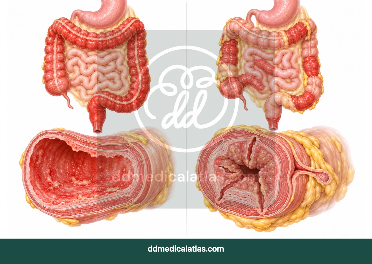
Ulcerative colitis versus Crohn's disease
A medical illustration comparing the continuous colonic inflammation associated with ulcerative colitis with the patchy, transmural inflammation that may occur in Crohn’s disease.
Public illustration library
Browse a selected collection of educational previews. Create a free account to open an illustration in the annotation workspace.
Gastroenterology
A medical illustration comparing the continuous colonic inflammation associated with ulcerative colitis with the patchy, transmural inflammation that may occur in Crohn’s disease.
Gastroenterology
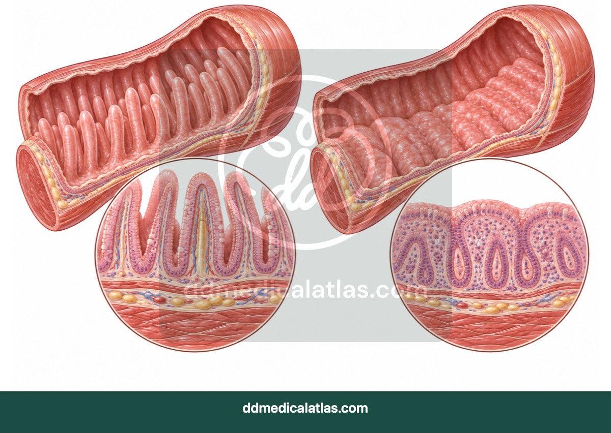
A medical illustration comparing healthy small-intestinal villi with the villous atrophy associated with untreated coeliac disease and reduced nutrient absorption.
Gastroenterology
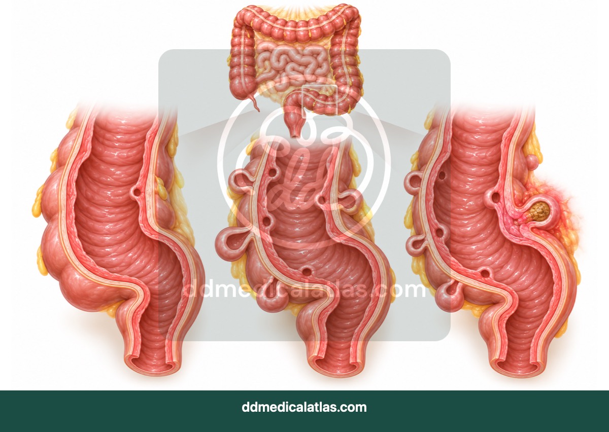
A medical illustration showing diverticula in the colon and the inflammation that distinguishes diverticulitis from uncomplicated diverticulosis.
Gastroenterology
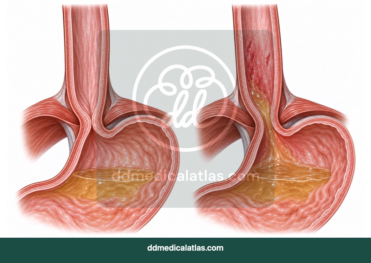
A medical illustration comparing a healthy lower oesophagus with irritation and inflammation caused by gastro-oesophageal reflux.
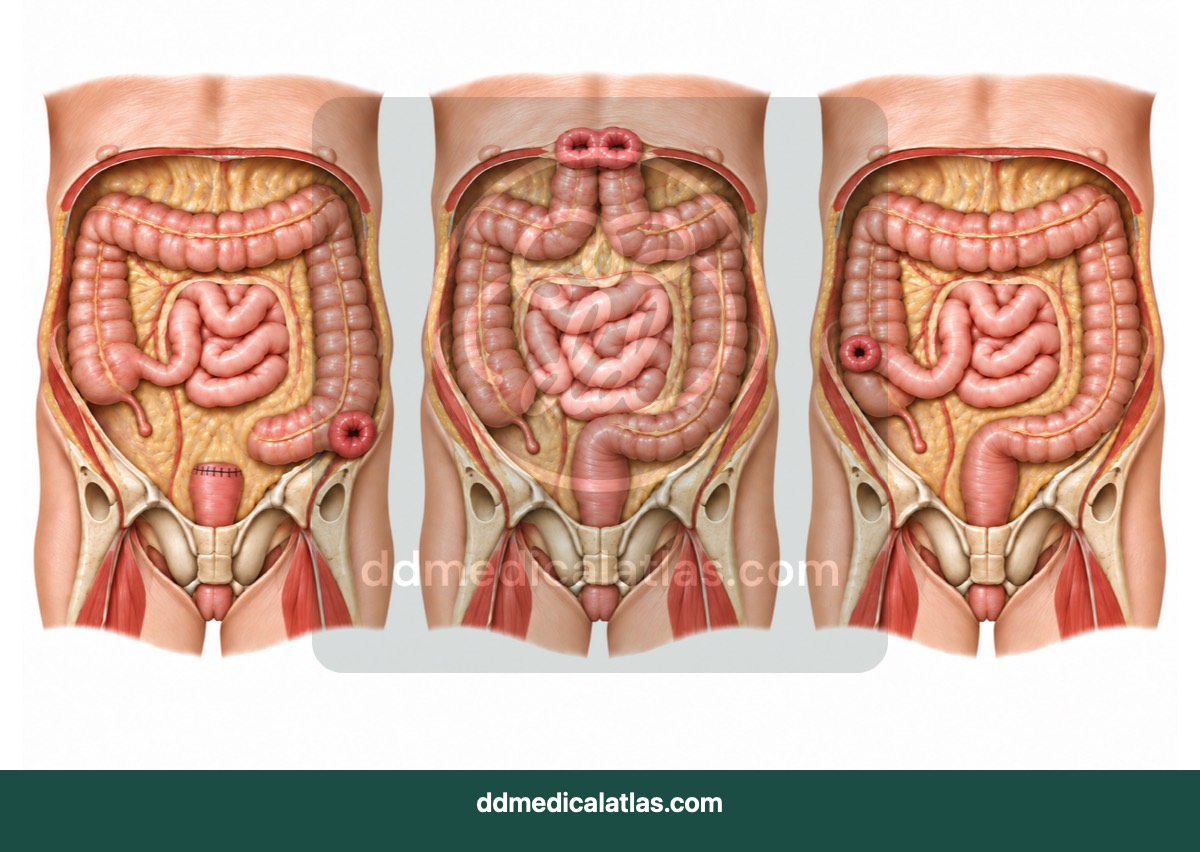
Gastroenterology
A medical illustration comparing common abdominal stoma configurations, including ileostomy and colostomy anatomy.
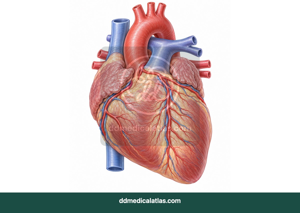
Cardiology
A labelled medical illustration of normal cardiac anatomy and the principal coronary arteries supplying the heart muscle.
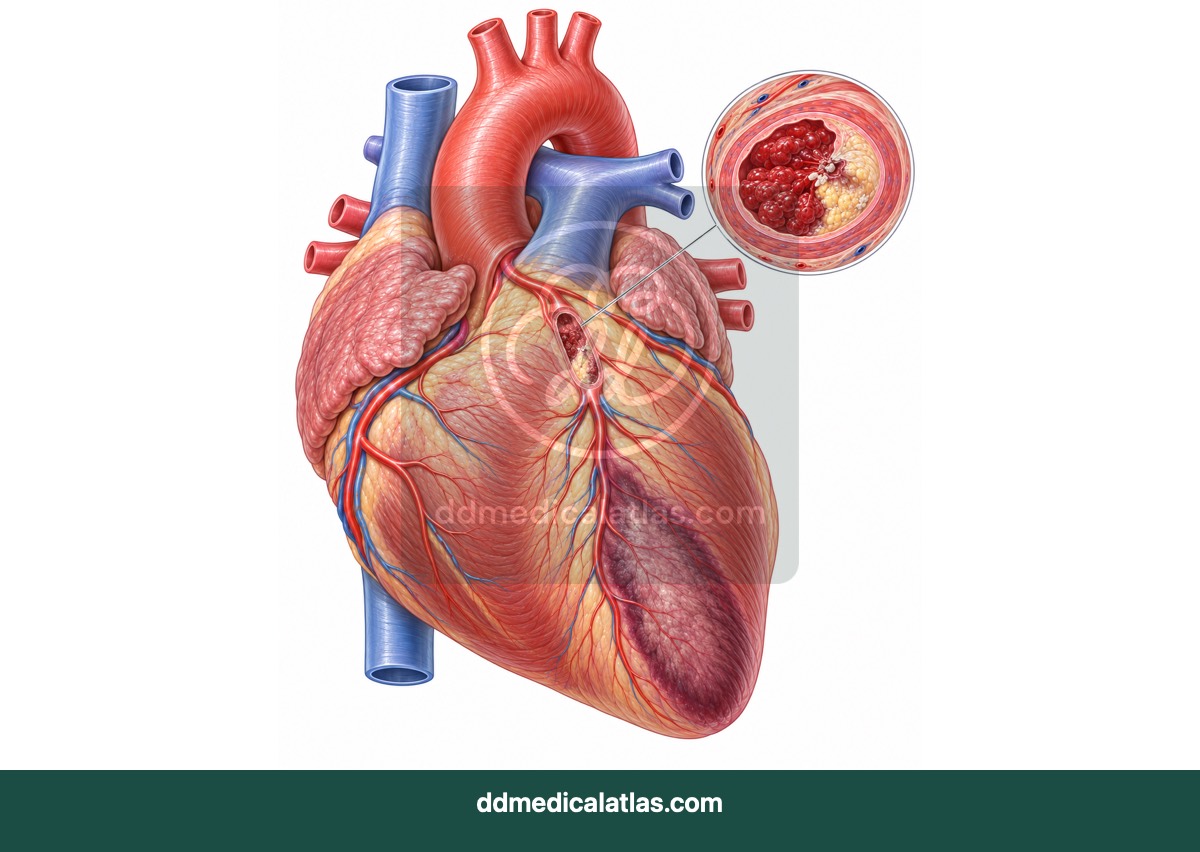
Cardiology
A medical illustration explaining how coronary artery narrowing and blockage can reduce blood supply and cause myocardial infarction.
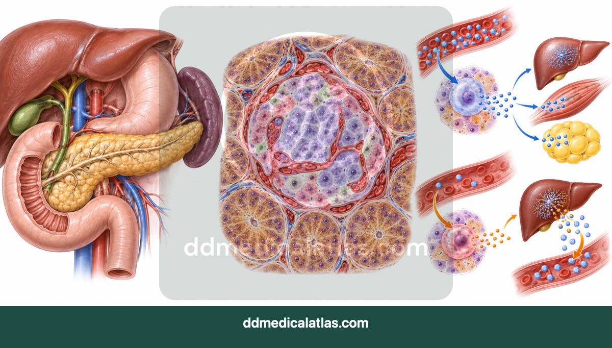
Endocrinology and Diabetes
A medical illustration of pancreatic islets and the roles of insulin and glucagon in regulating blood glucose.
Respiratory
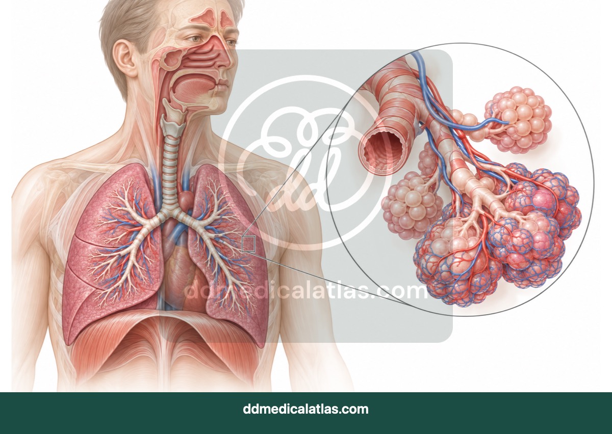
A medical illustration of the respiratory tract, lungs and branching airways used in breathing and gas exchange.
Respiratory
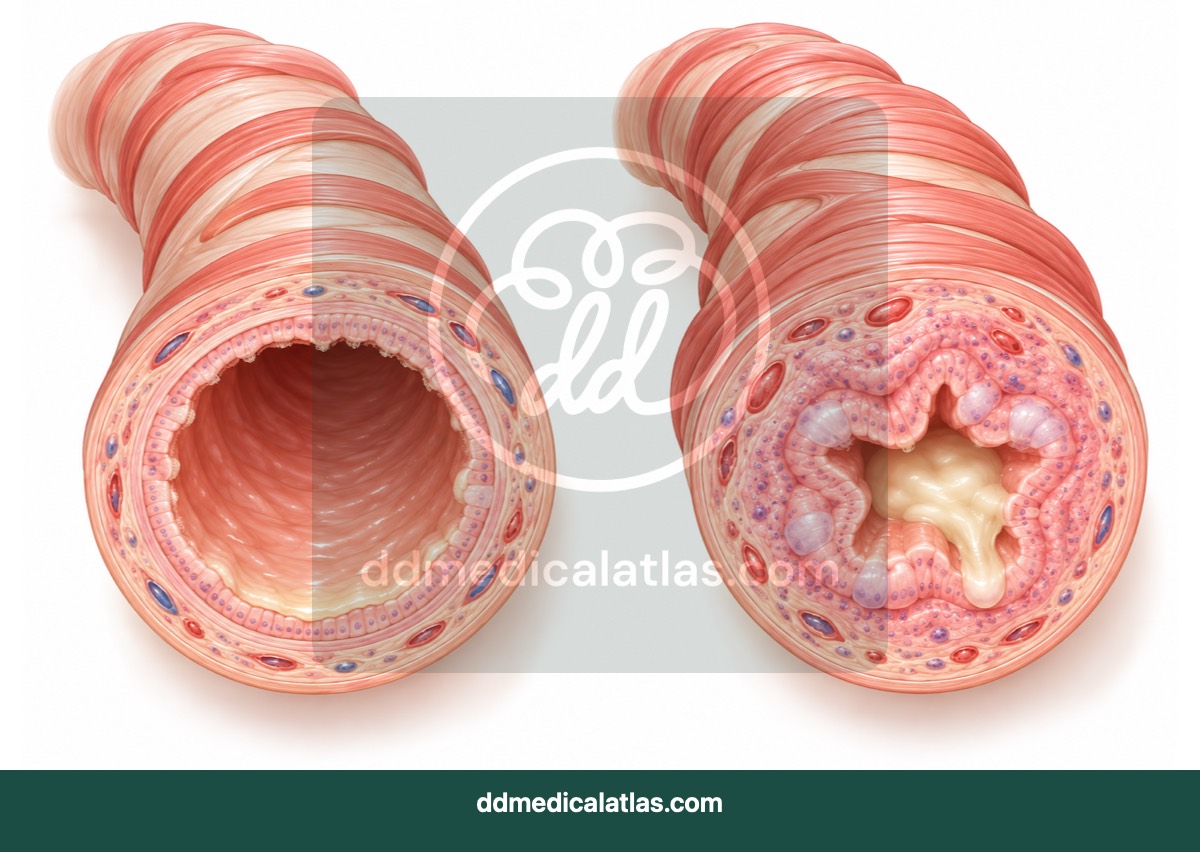
A cross-sectional medical illustration comparing a normal bronchiole with airway narrowing, inflammation and mucus during asthma.
Respiratory
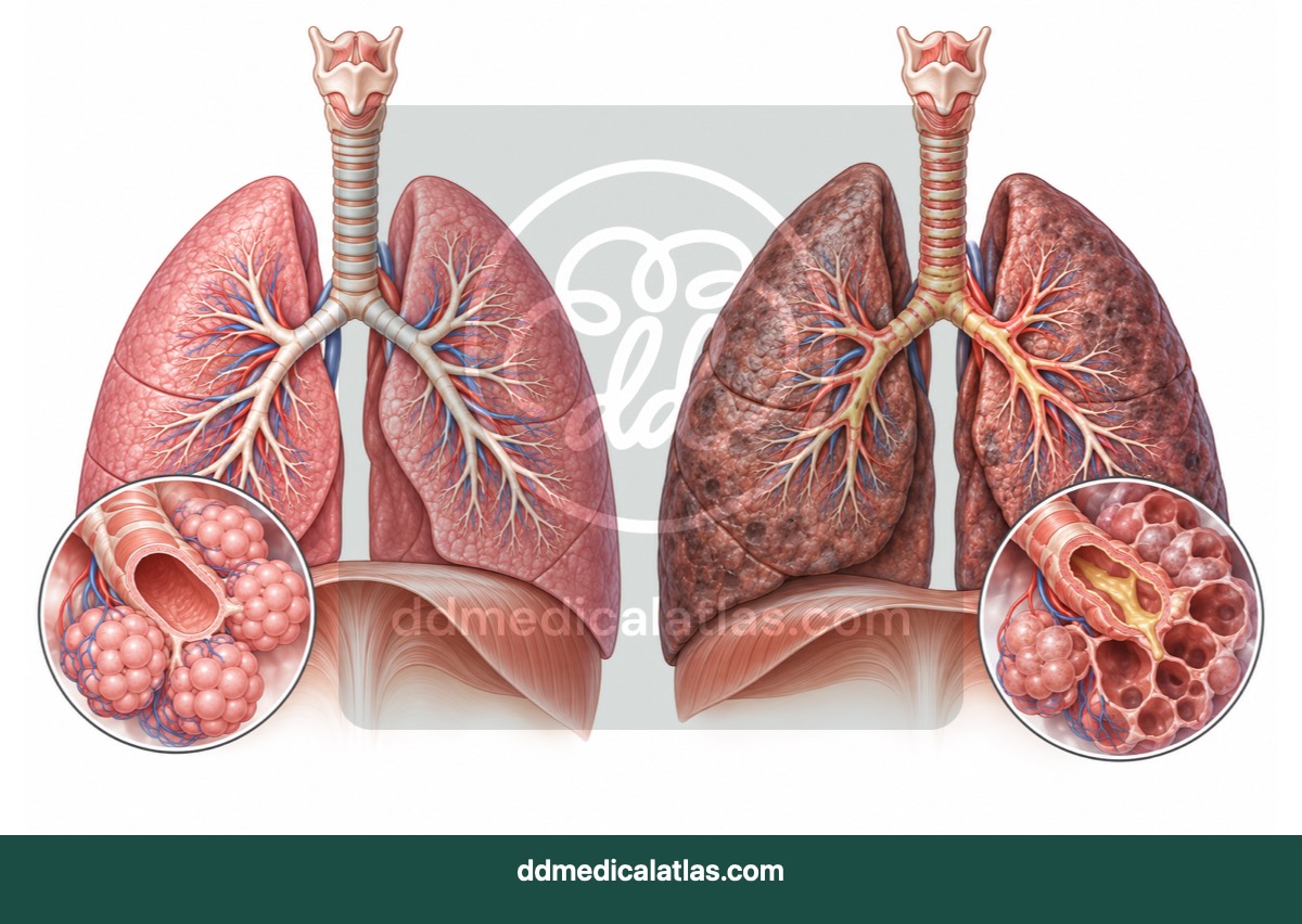
A medical illustration summarising airway obstruction and lung changes associated with chronic obstructive pulmonary disease.
Musculoskeletal
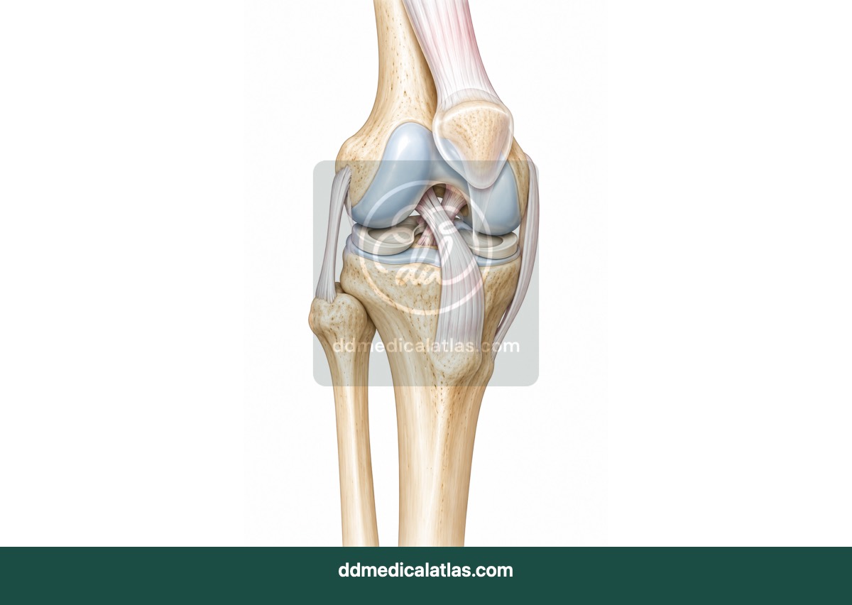
A medical illustration of normal knee anatomy, including the principal bones, cartilage, menisci and supporting ligaments.
Musculoskeletal
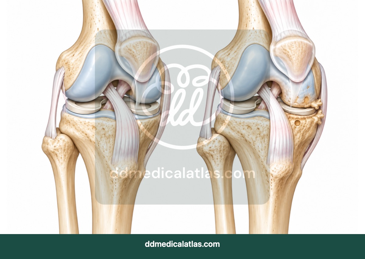
A medical illustration comparing a healthy knee joint with cartilage loss, joint-space narrowing and bony changes in osteoarthritis.
Musculoskeletal
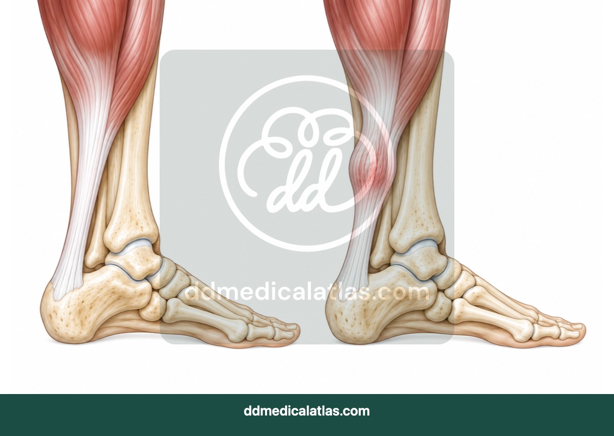
A medical illustration showing the Achilles tendon and common sites of insertional and mid-portion tendinopathy.
Podiatry
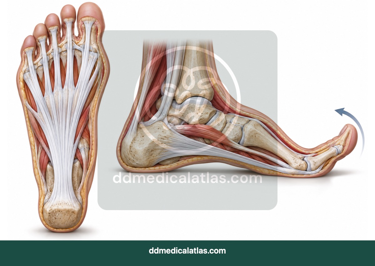
A plantar foot illustration showing the origin, course and insertion of the plantar fascia and its role in the windlass mechanism.
Podiatry
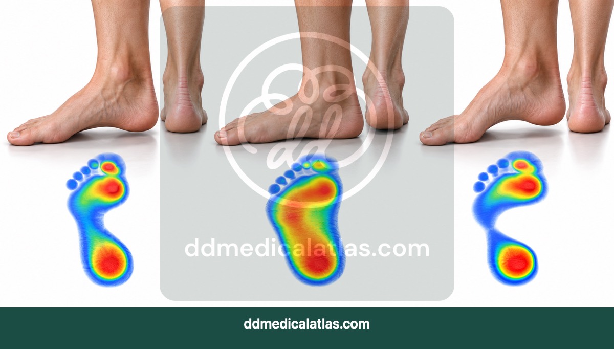
A comparative medical illustration of a normal foot arch, pes planus and pes cavus for podiatry and gait education.
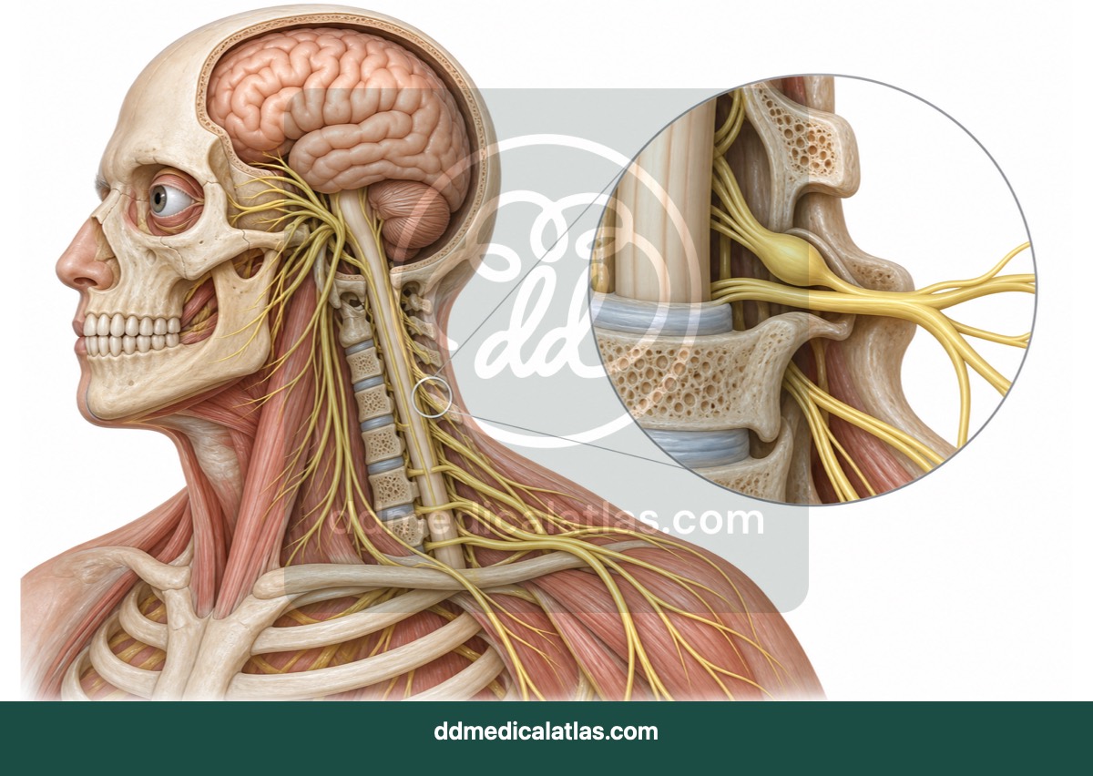
Neurology
A medical illustration providing an overview of the brain, skull, spinal cord and major peripheral nerve pathways.
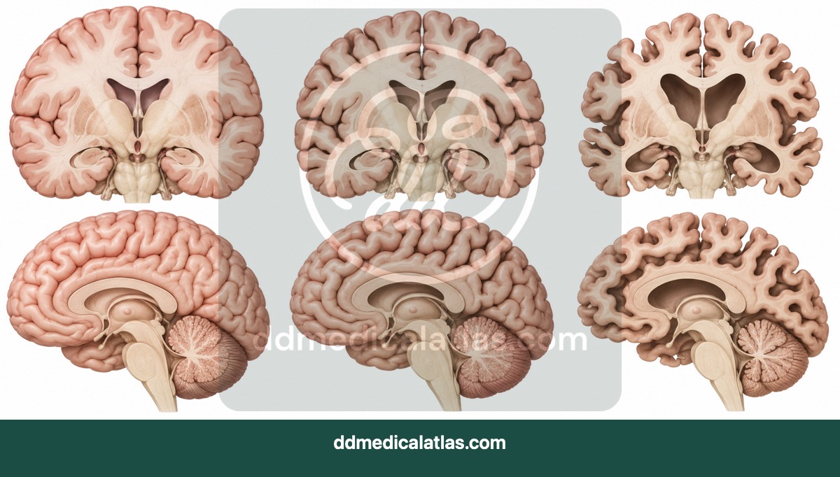
Geriatric
A medical illustration comparing a healthy younger brain, expected age-related changes and the more pronounced cortical atrophy associated with Alzheimer’s disease.
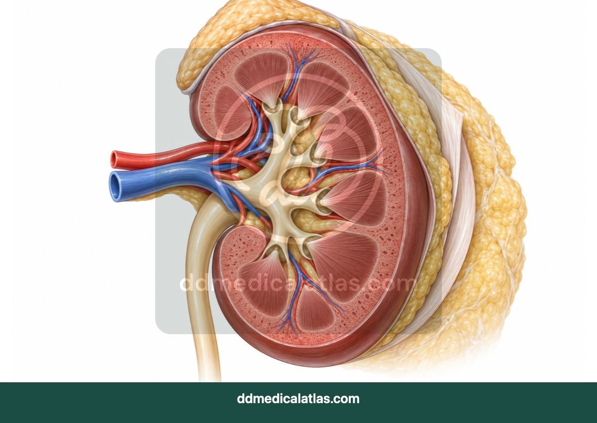
Renal and urinary
A cutaway medical illustration showing the cortex, medulla, pyramids, calyces and renal pelvis of the kidney.
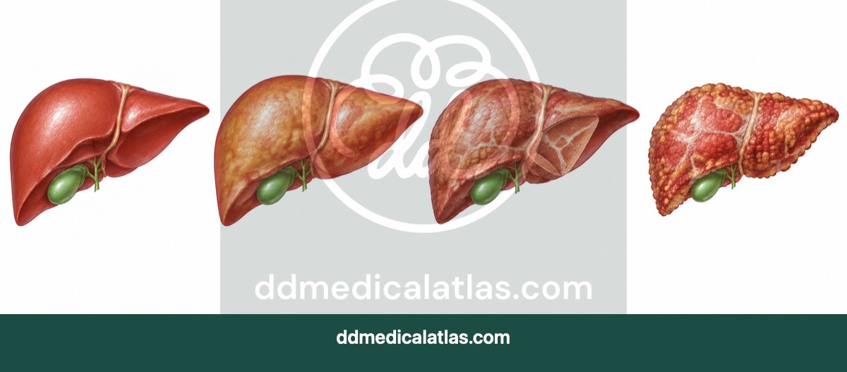
Gastroenterology
A medical illustration showing the progression from a healthy liver through fibrosis to cirrhosis in chronic liver disease.
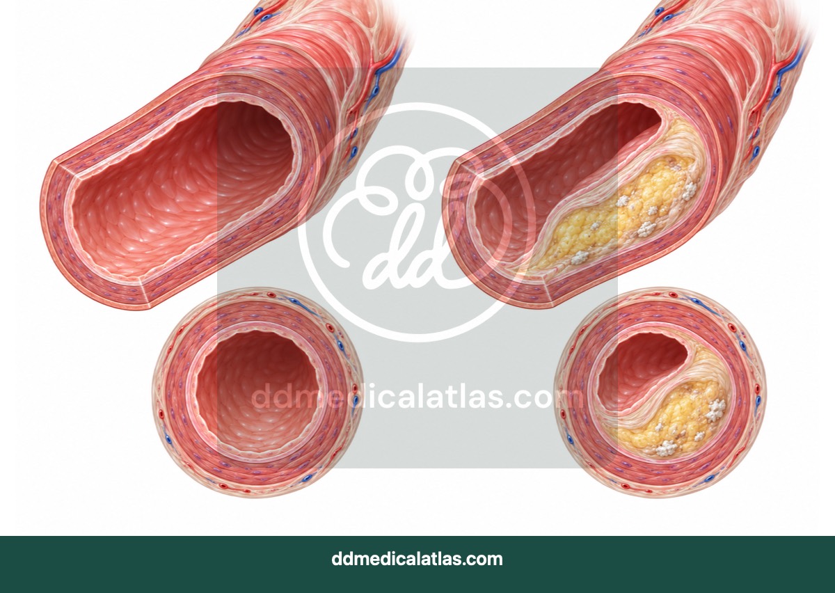
Cardiology
A medical illustration comparing a healthy artery with atherosclerotic plaque formation, arterial narrowing and restricted blood flow.
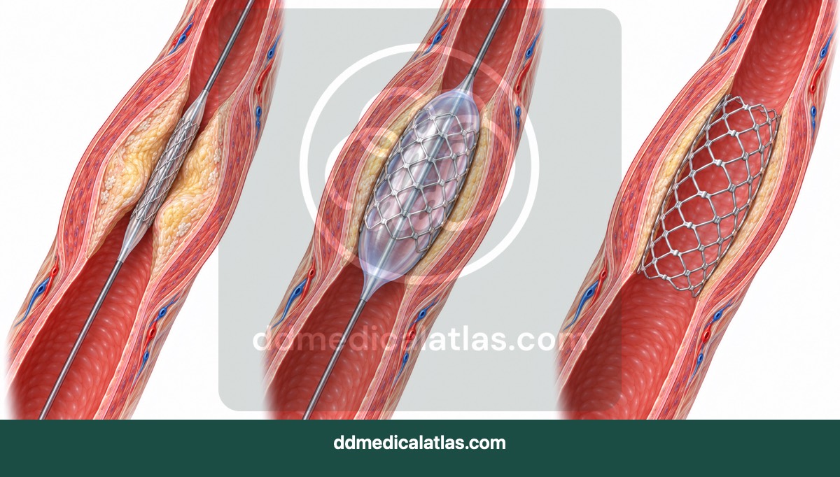
Cardiology
A step-by-step medical illustration showing balloon angioplasty and coronary stent placement used to reopen a narrowed coronary artery.
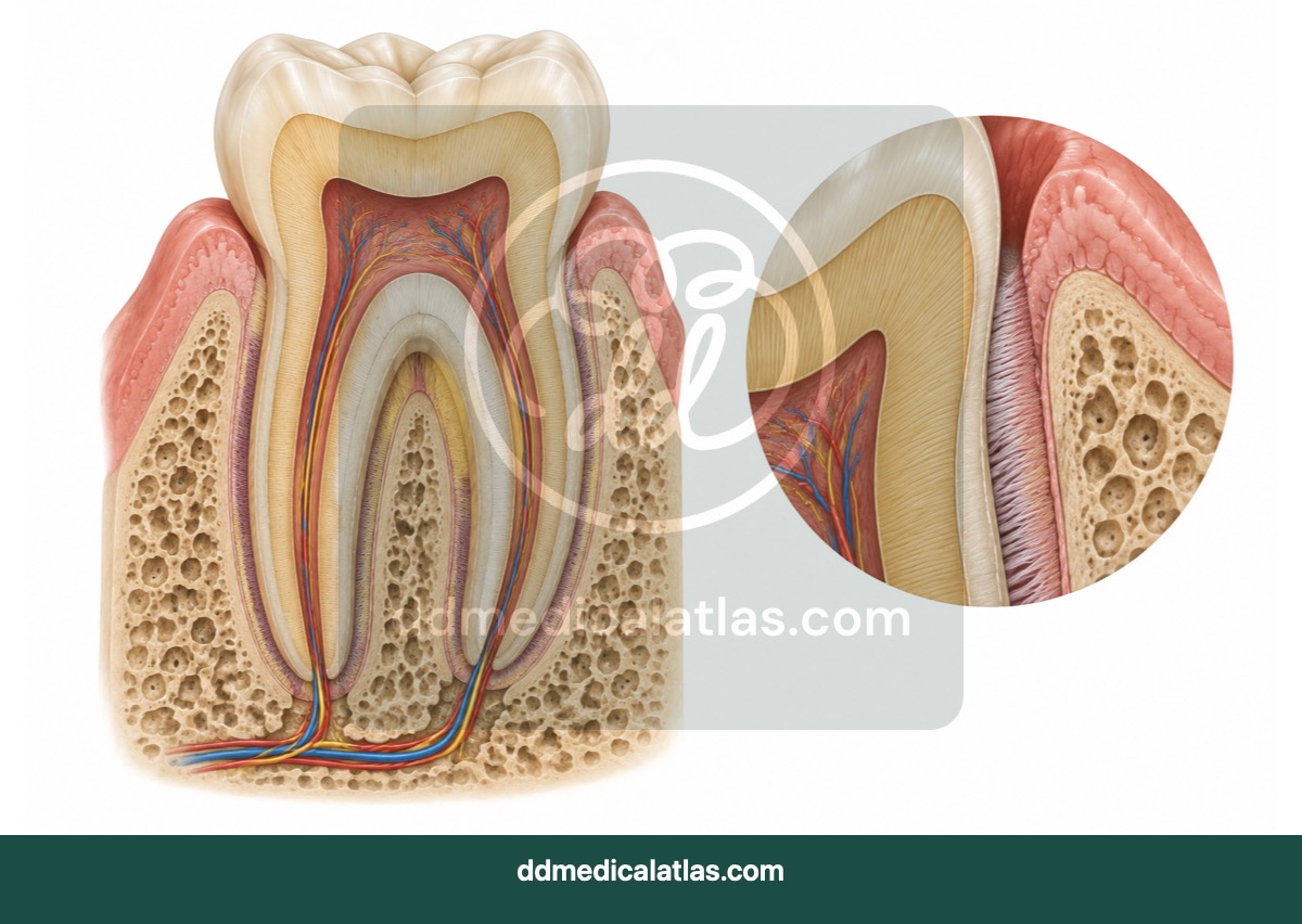
Dentistry
A labelled dental illustration showing the crown, enamel, dentine, pulp, roots and supporting periodontal tissues of a tooth.
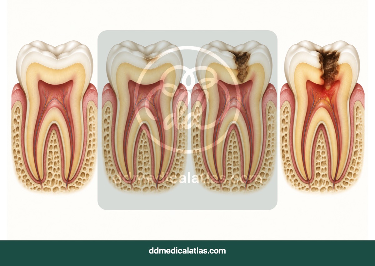
Dentistry
A medical illustration showing the progression of dental caries from early enamel decay through dentine involvement to the dental pulp.
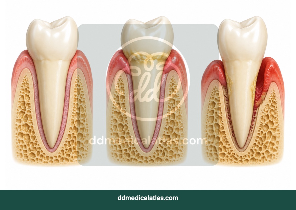
Dentistry
A comparative dental illustration showing healthy gums, gingivitis and progressive periodontal disease with loss of supporting tissue and bone.
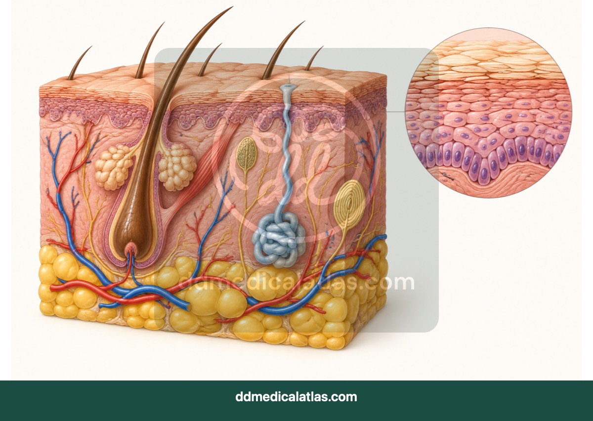
Dermatology
A cutaway medical illustration of normal skin showing the epidermis, dermis, subcutaneous tissue, hair follicles, glands, nerves and vessels.
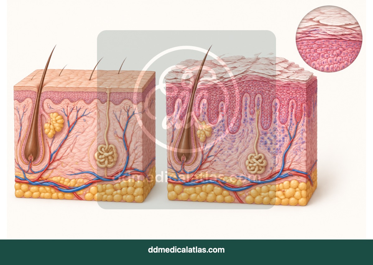
Dermatology
A medical illustration comparing healthy skin with the thickened epidermis, inflammation and accelerated skin-cell turnover associated with psoriasis.
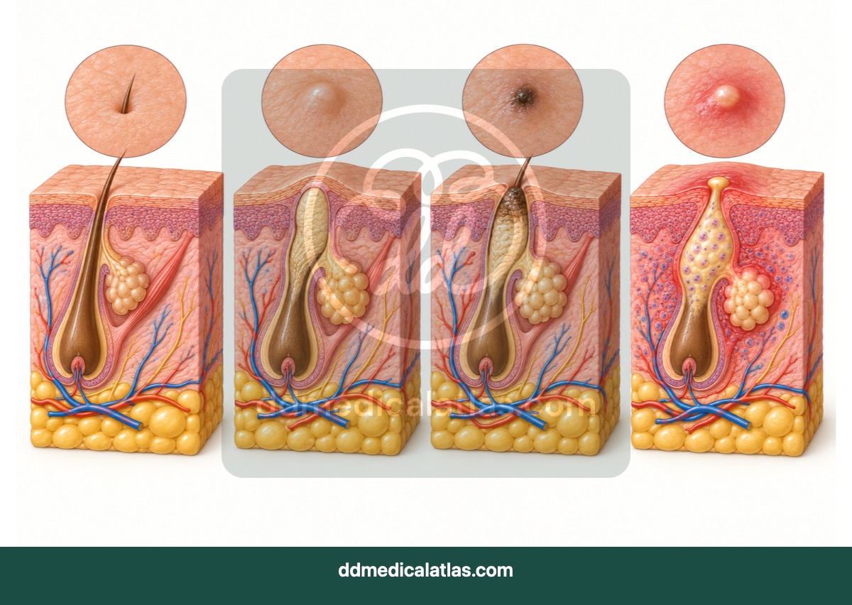
Dermatology
A medical illustration showing how blocked pilosebaceous units may progress through comedonal and inflammatory stages of acne vulgaris.
Dermatology
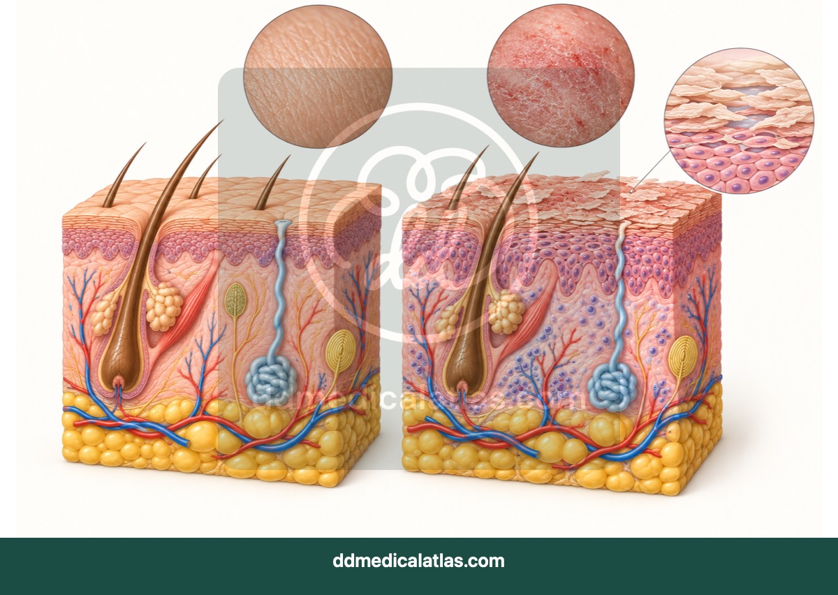
A comparative medical illustration showing healthy skin and the impaired skin barrier, dryness and inflammation associated with atopic dermatitis.
Dermatology
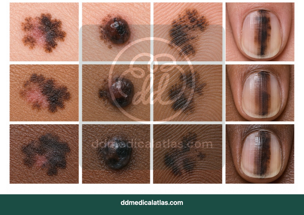
A clinical comparison illustrating varied melanoma appearances across different skin tones and anatomical sites for educational awareness.
Endocrinology and Diabetes
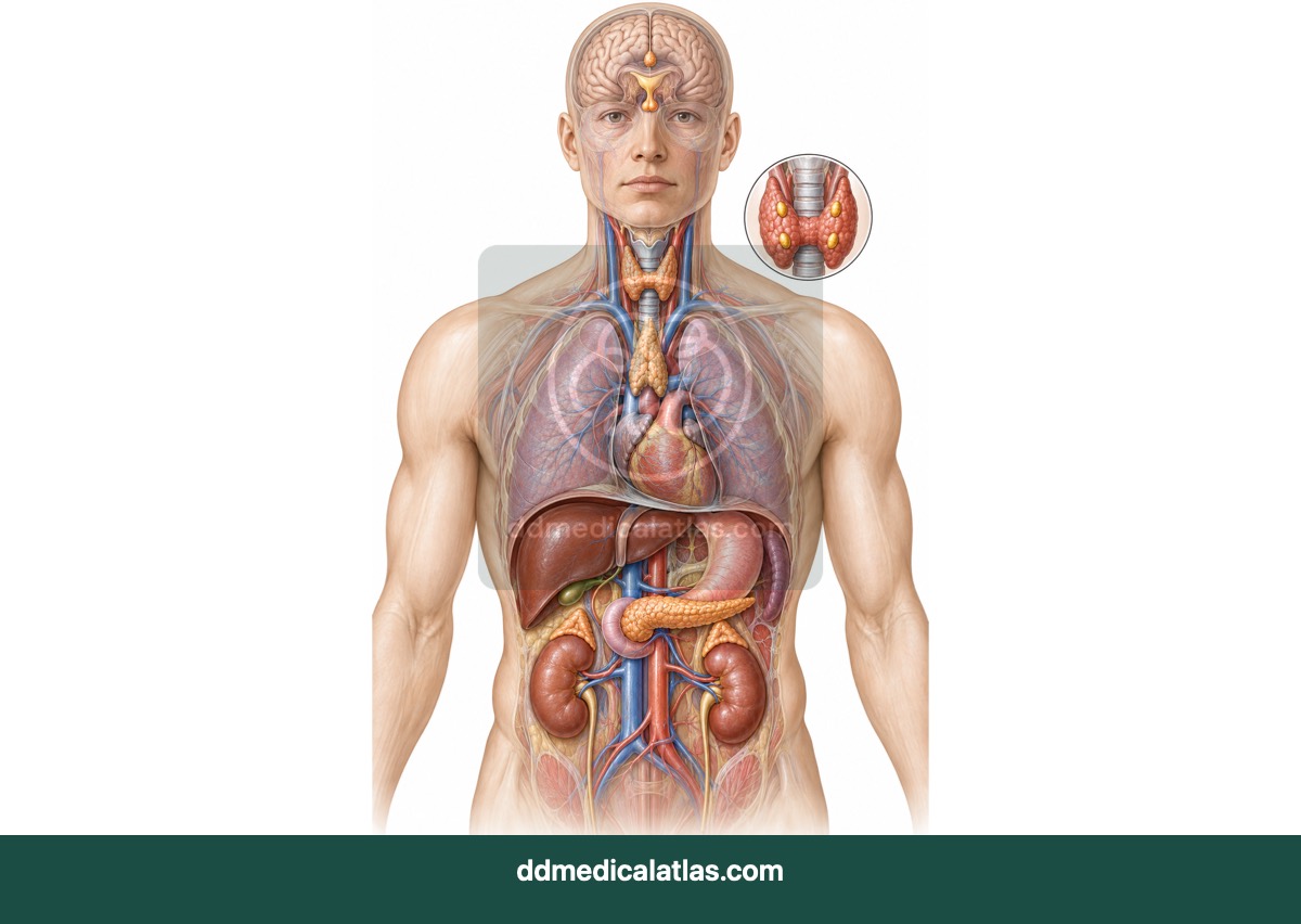
A medical illustration locating the principal endocrine glands and showing their roles in hormonal regulation across the body.
Endocrinology and Diabetes
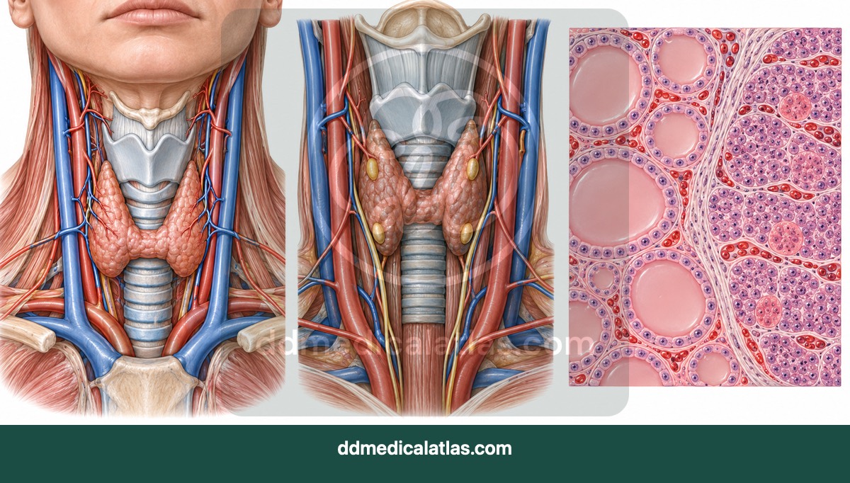
A medical illustration showing the position and anatomy of the thyroid and parathyroid glands in the neck.
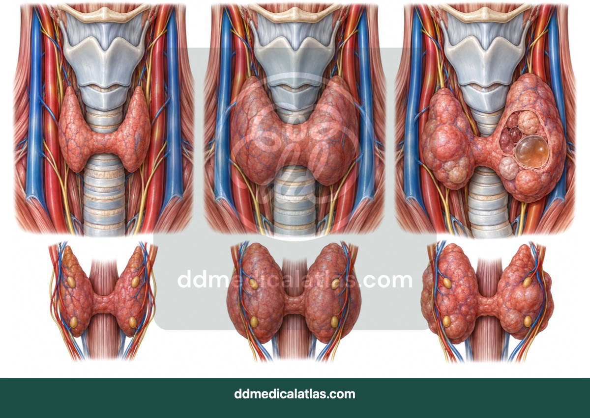
Endocrinology and Diabetes
A medical illustration comparing normal thyroid anatomy with thyroid enlargement and common patterns of thyroid nodules.
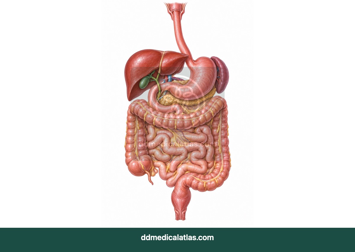
Gastroenterology
A medical illustration of the complete gastrointestinal tract and associated digestive organs from the oesophagus to the rectum.
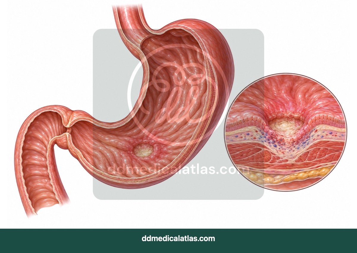
Gastroenterology
A cutaway medical illustration showing a gastric ulcer extending through the protective lining of the stomach.
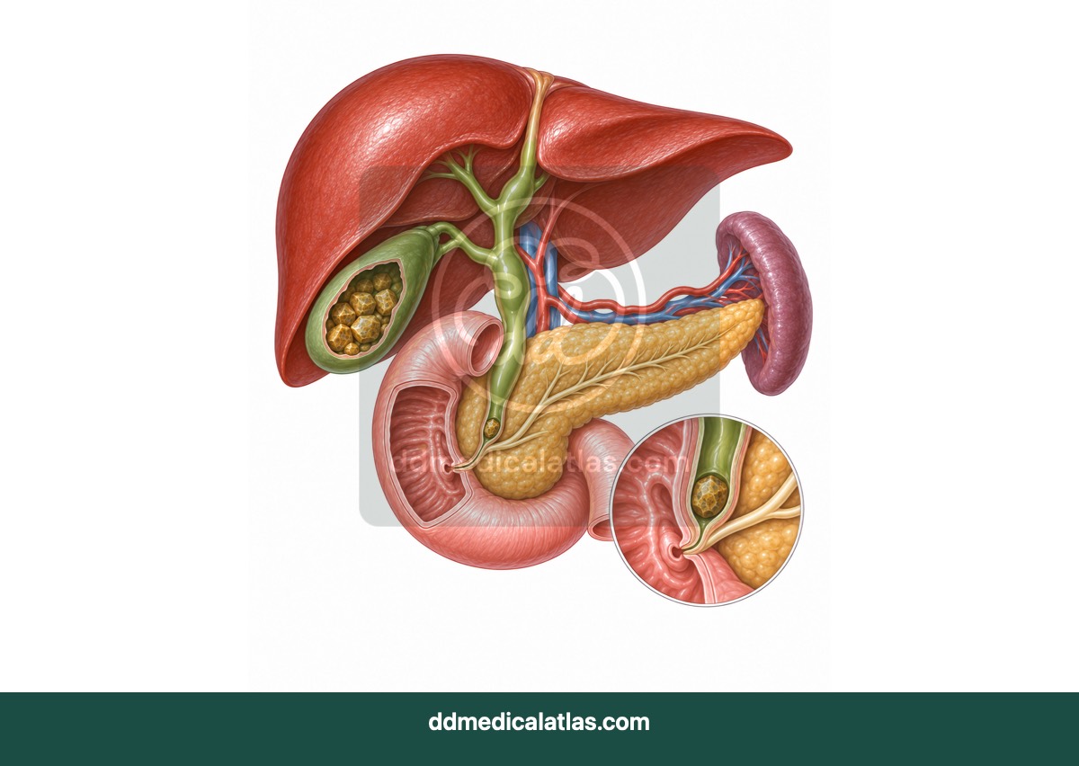
Gastroenterology
A medical illustration showing gallstones within the gallbladder and bile duct obstruction affecting normal bile drainage.
Gastroenterology
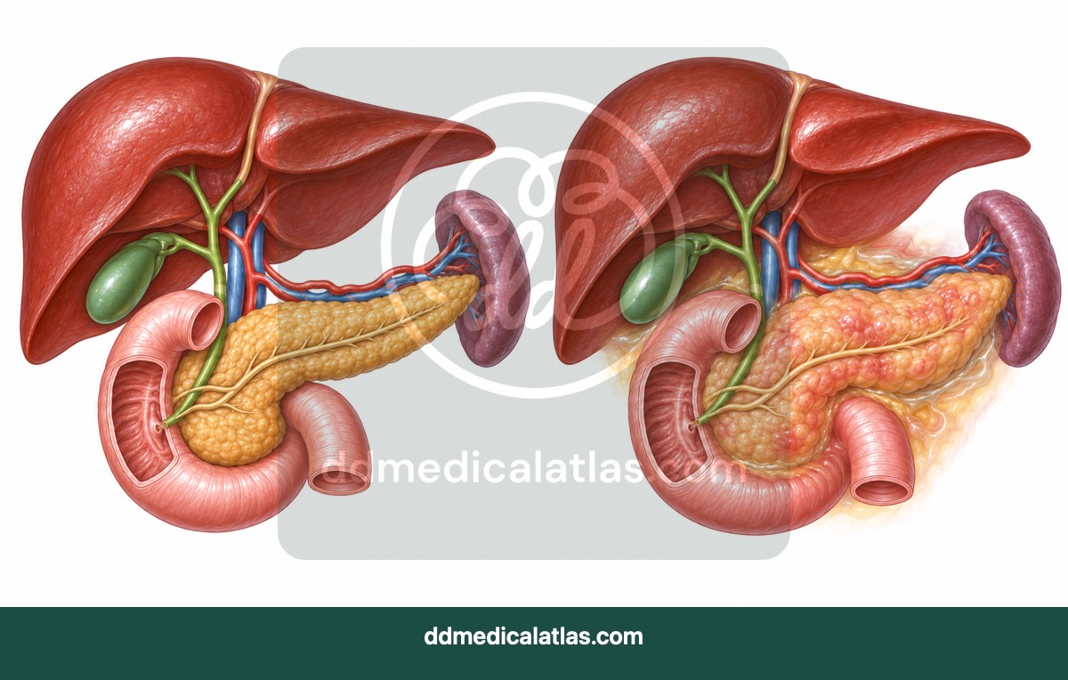
A medical illustration showing pancreatic inflammation and surrounding anatomical structures in acute pancreatitis.
Gastroenterology
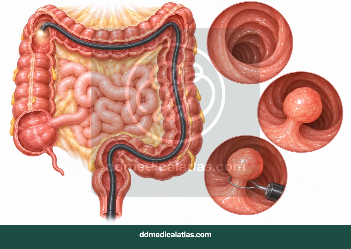
A medical illustration showing colonoscope navigation through the large bowel and endoscopic removal of a colonic polyp.
Gastroenterology
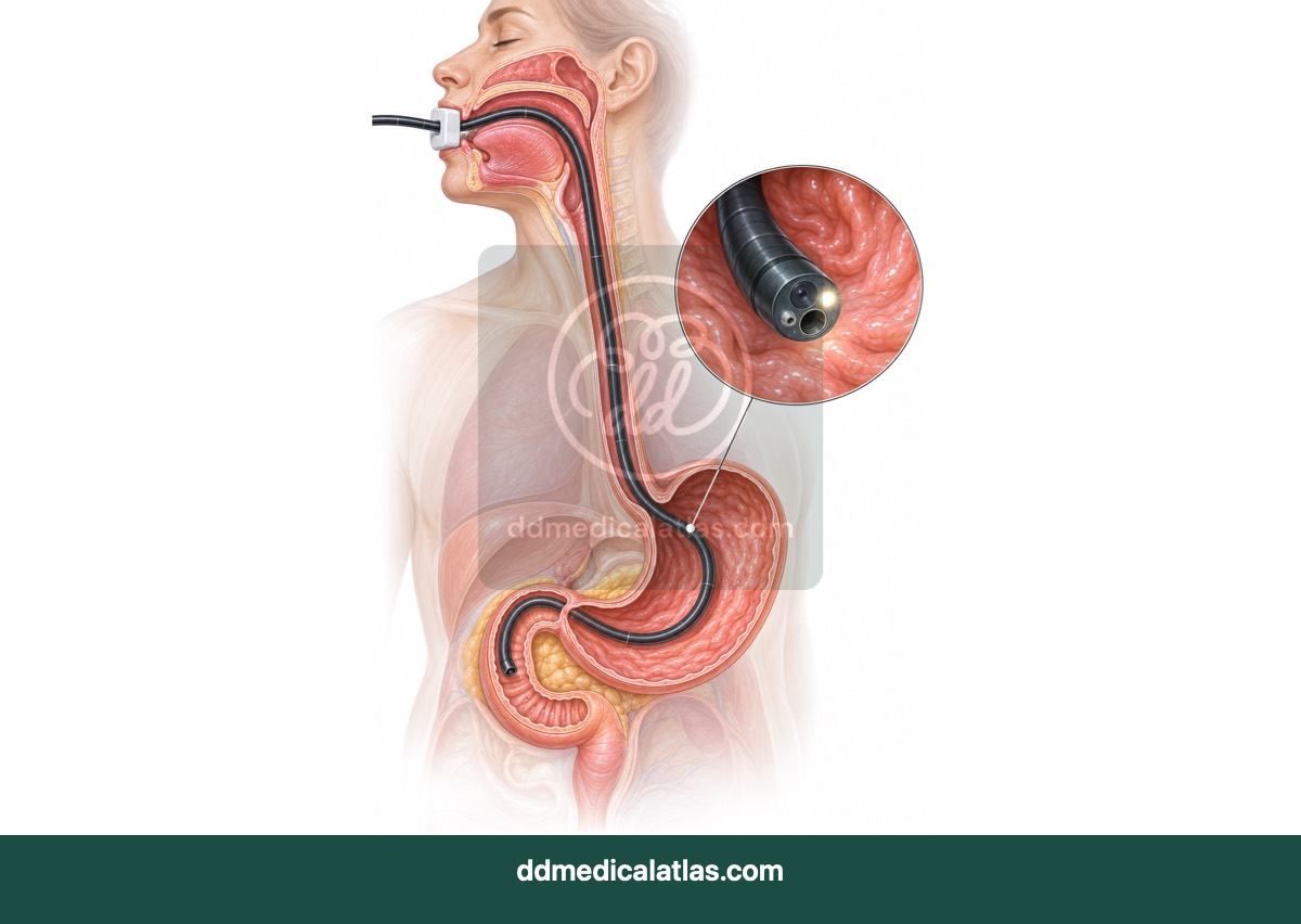
A medical illustration showing the route of an upper gastrointestinal endoscope through the oesophagus, stomach and duodenum.
Gastroenterology
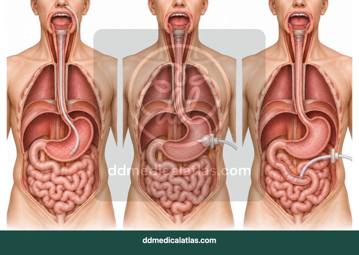
A medical illustration explaining enteral nutrition support and common tube-feeding routes into the gastrointestinal tract.
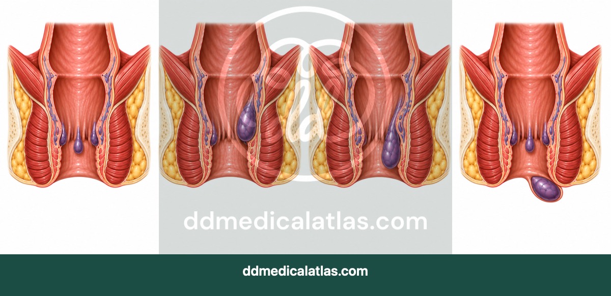
Gastroenterology
A medical illustration showing internal and external haemorrhoids and the anatomical features used in clinical classification.
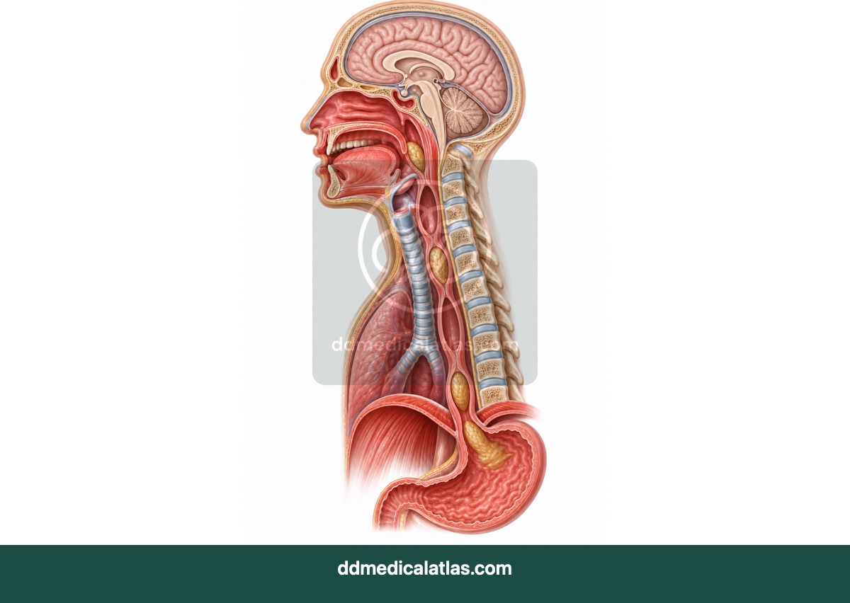
Gastroenterology
A medical illustration showing the coordinated stages of swallowing and movement of a food bolus through the oesophagus.
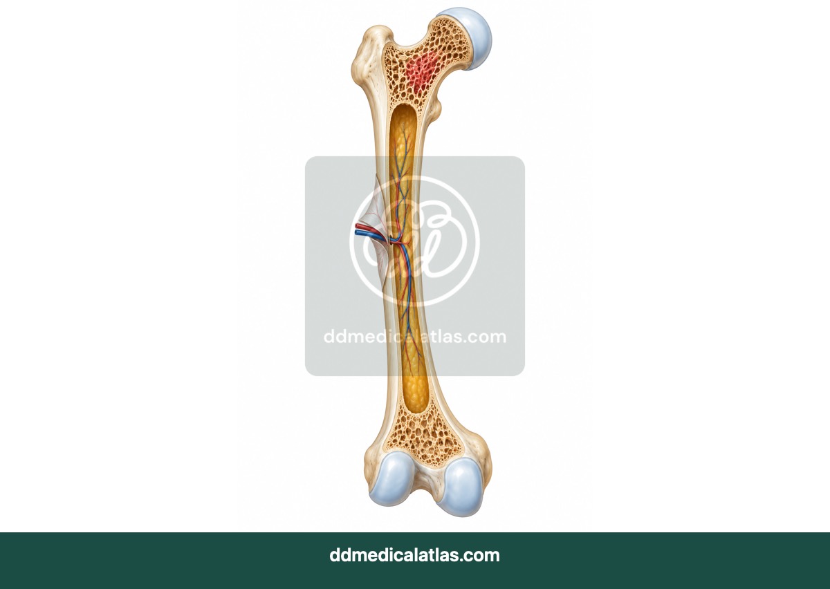
Musculoskeletal
A cutaway medical illustration of long-bone structure showing cortical bone, trabecular bone, marrow and growth regions.
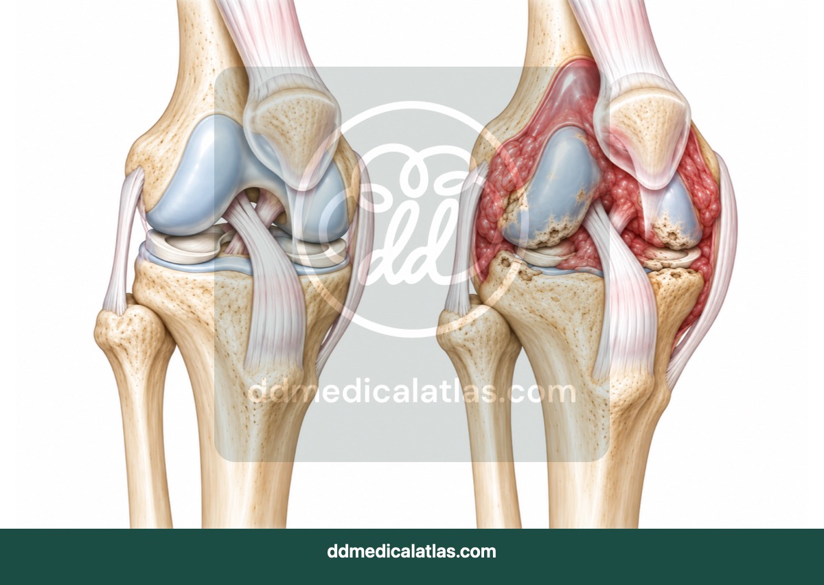
Musculoskeletal
A medical illustration comparing a healthy knee with synovial inflammation and joint damage associated with rheumatoid arthritis.
Musculoskeletal
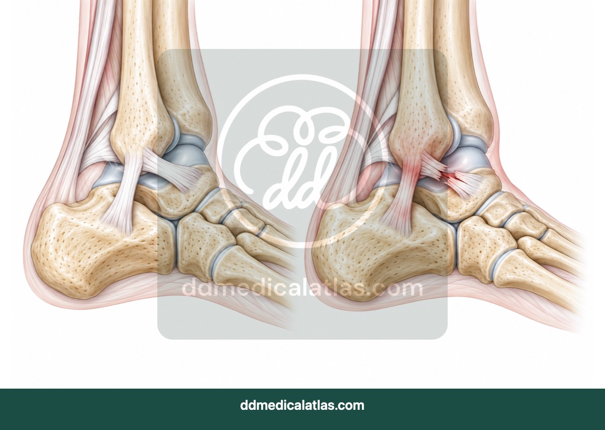
A medical illustration showing the lateral ankle ligaments and common patterns of injury during an ankle sprain.
Musculoskeletal
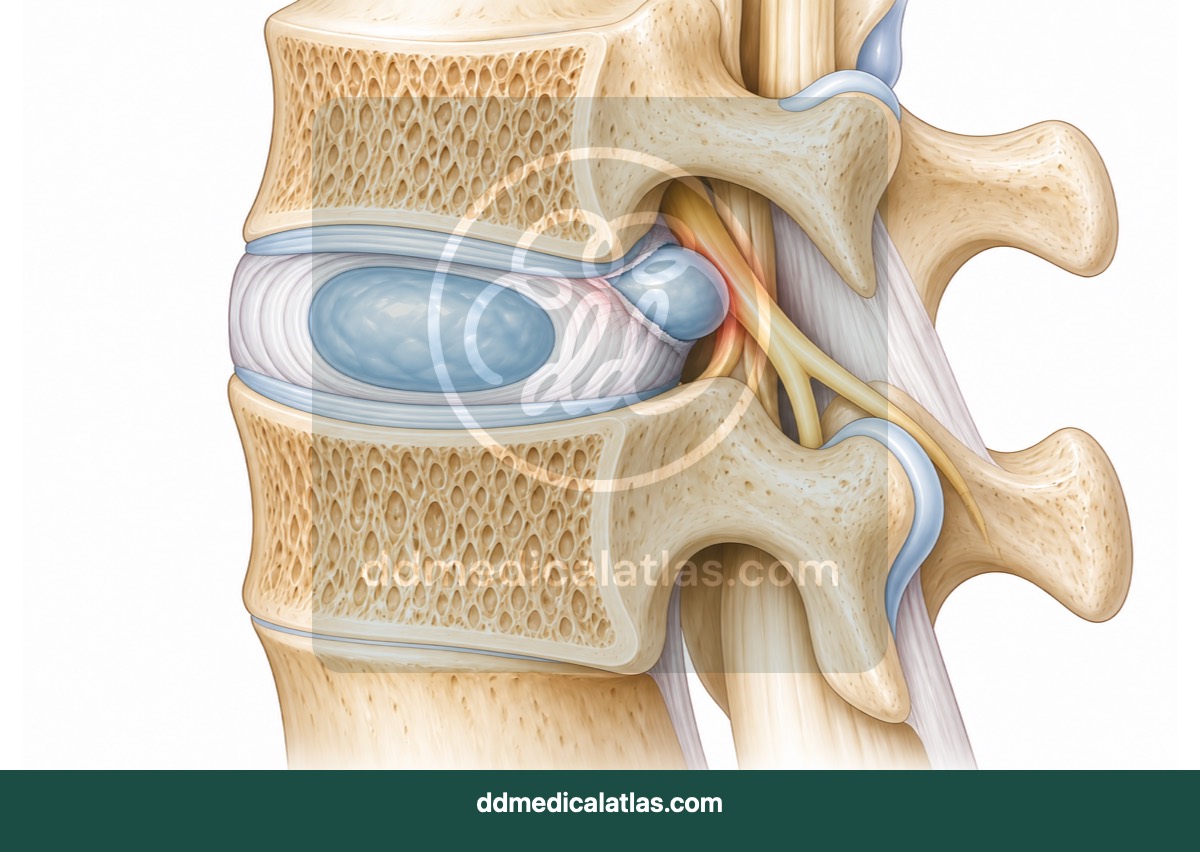
A spinal cutaway illustration showing an intervertebral disc herniation and its relationship to adjacent nerve roots.
Musculoskeletal
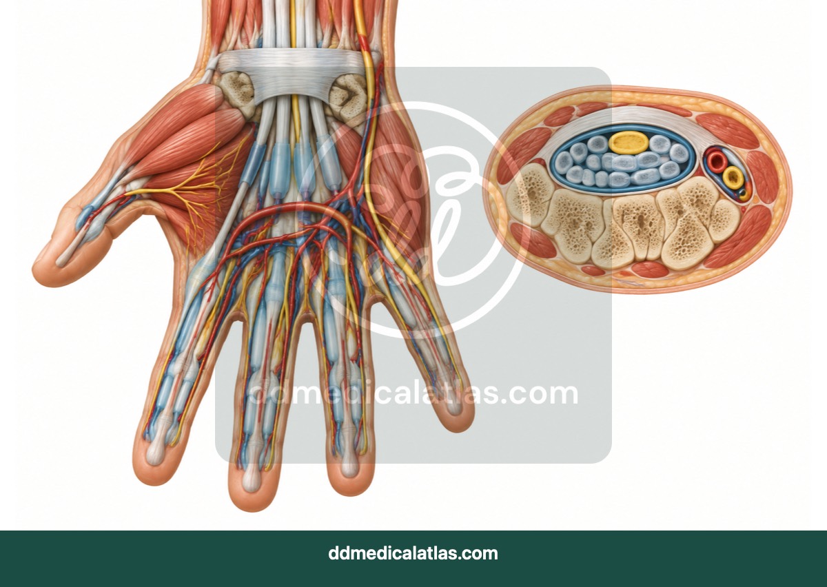
A layered medical illustration showing the carpal tunnel, median nerve, flexor tendons and surrounding wrist anatomy.
Neurology
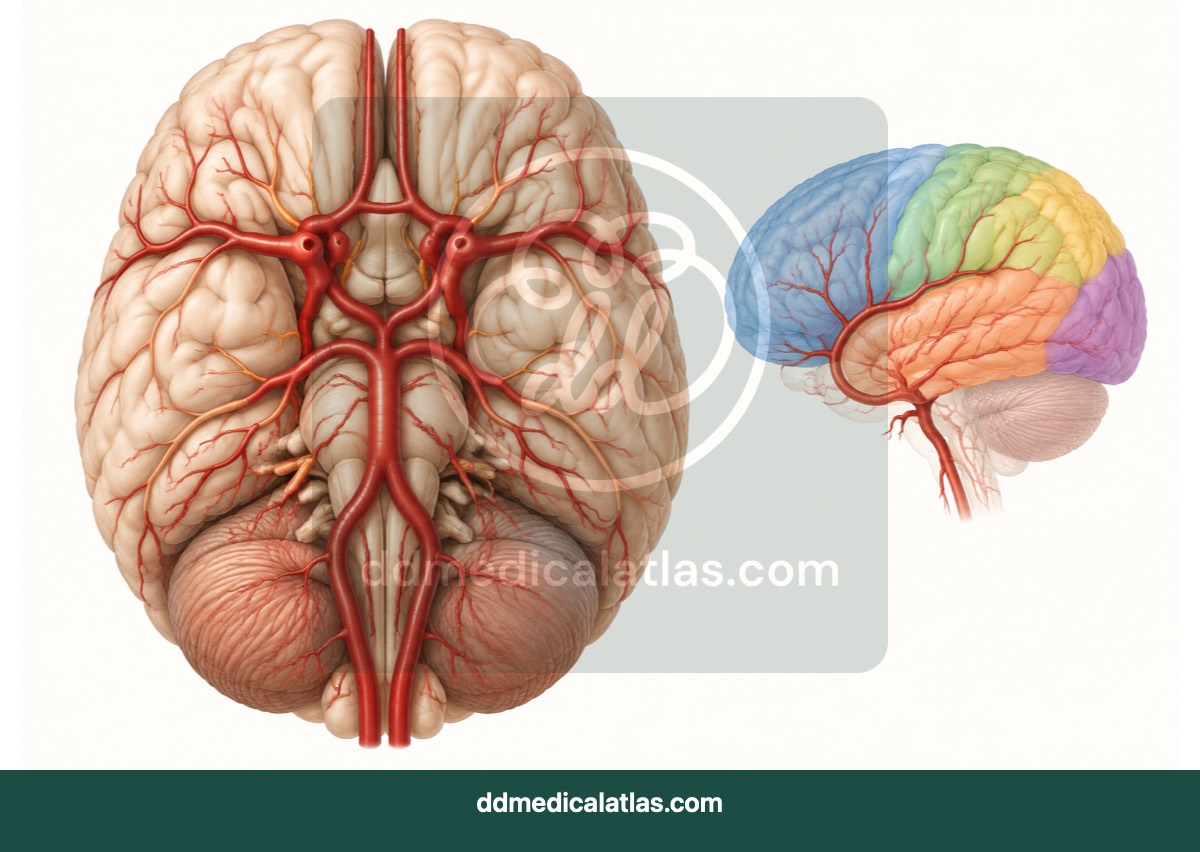
A medical illustration of cerebral arterial circulation highlighting the major vessels and communicating arteries of the Circle of Willis.
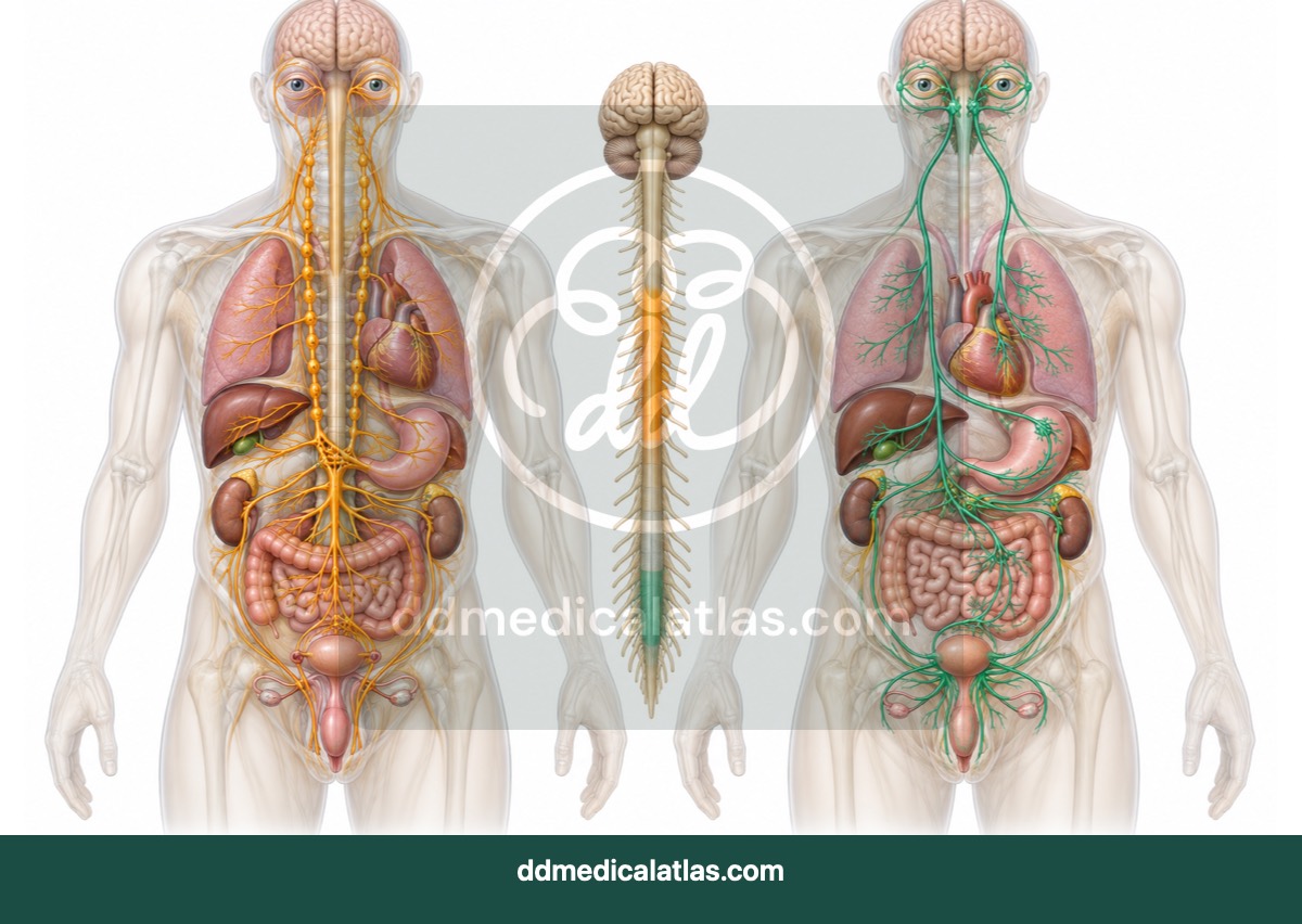
Neurology
A medical illustration comparing sympathetic and parasympathetic pathways and their effects on major organs.
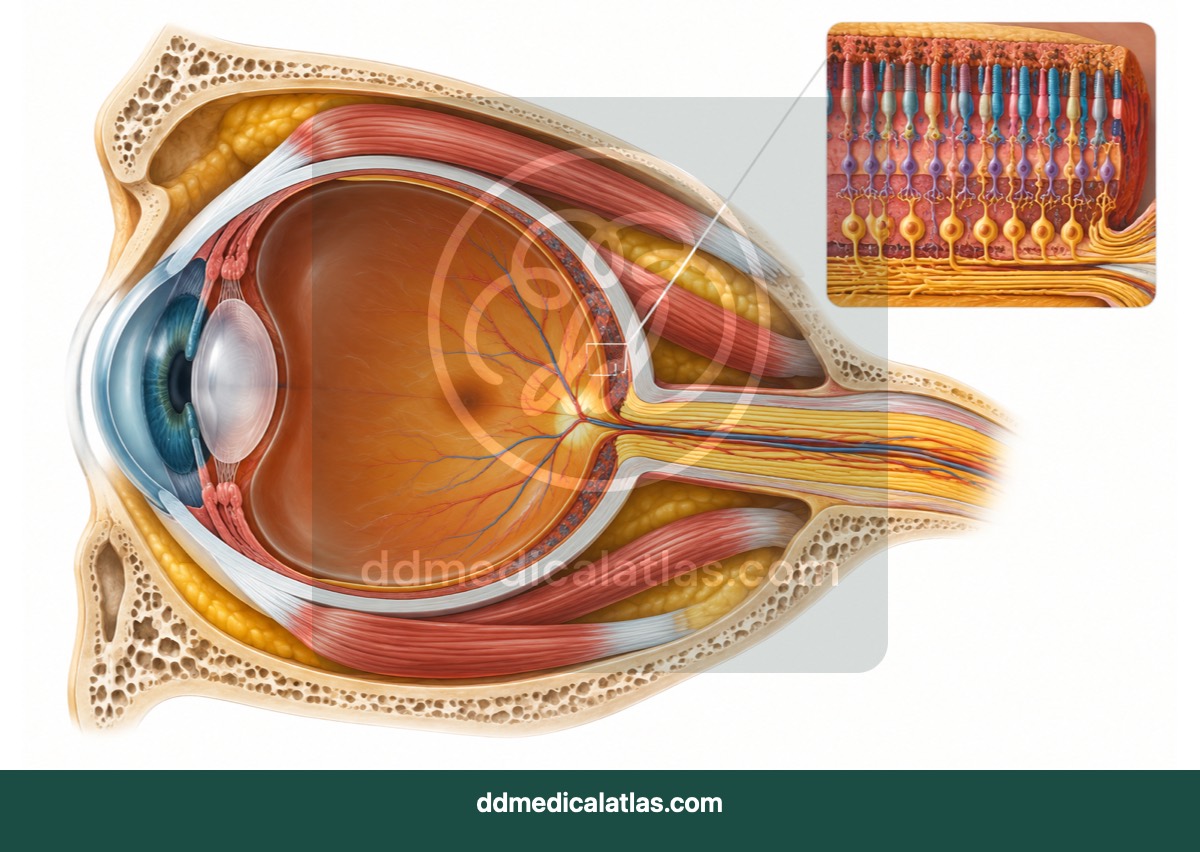
Ophthalmology
A cutaway medical illustration showing the structures of the eye, optic nerve, extraocular muscles and surrounding orbit.
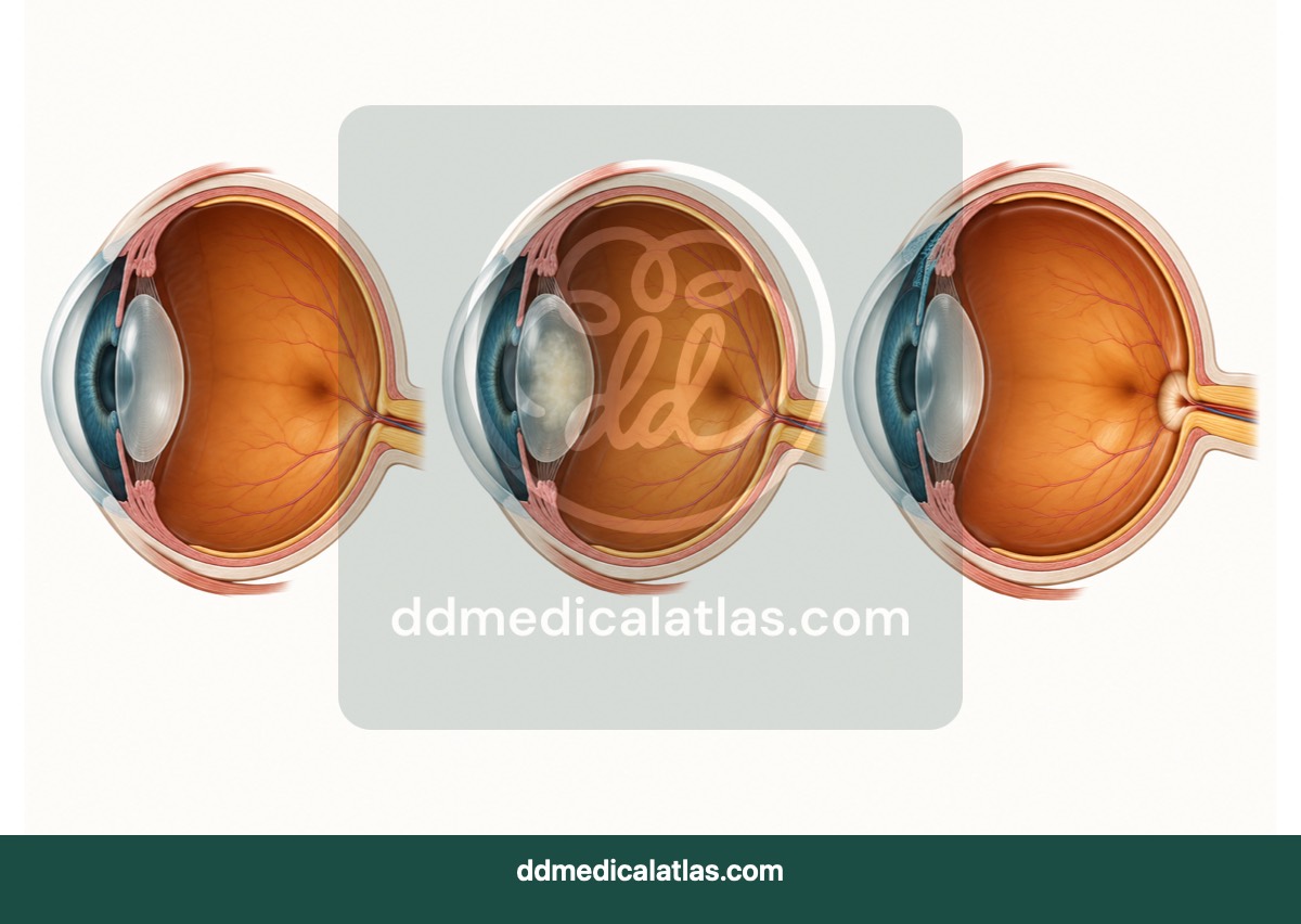
Ophthalmology
A comparative medical illustration showing lens clouding in cataract and pressure-related optic-nerve damage in glaucoma.
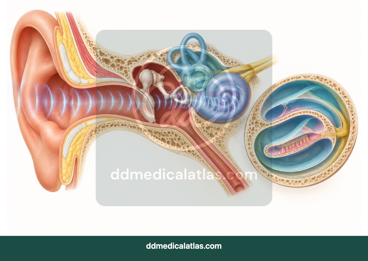
ENT — Otolaryngology
A medical illustration showing external, middle and inner ear anatomy and the pathway of sound conduction.
ENT — Otolaryngology
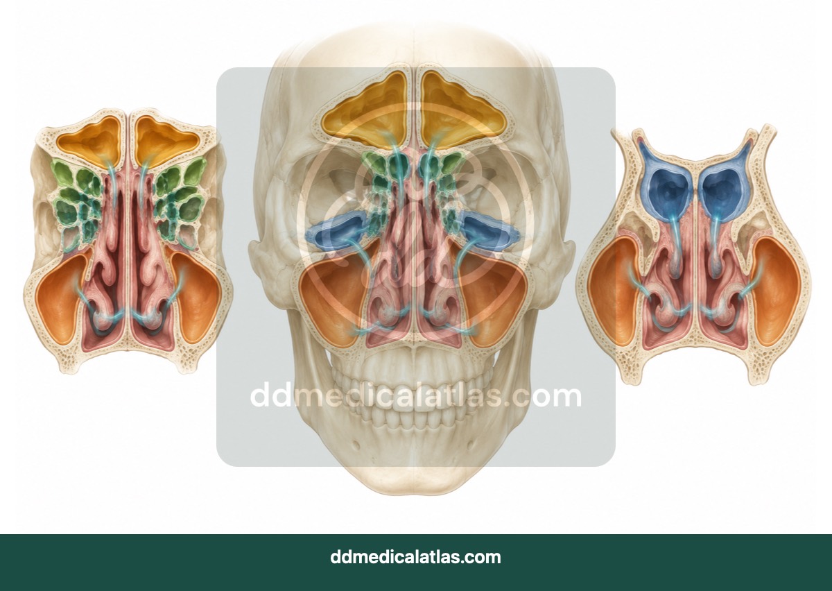
A facial cutaway illustration showing the frontal, ethmoid, sphenoid and maxillary sinuses and their drainage pathways.
Renal and urinary
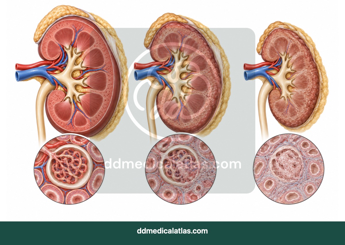
A medical illustration showing structural and functional changes as chronic kidney disease progresses towards kidney failure.
Renal and urinary
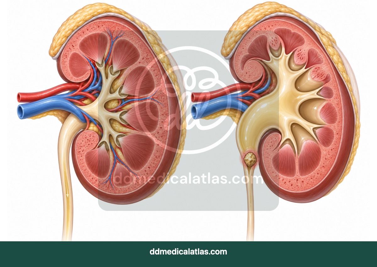
A medical illustration showing an obstructing urinary stone and the resulting dilation of the renal collecting system in hydronephrosis.
Renal and urinary
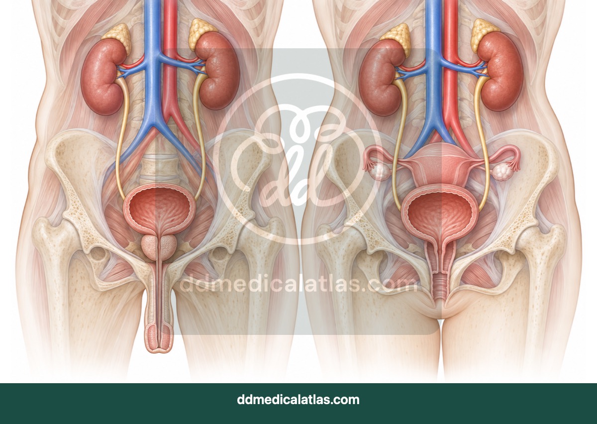
A comparative medical illustration showing male and female urinary tract anatomy, including the kidneys, ureters, bladder and urethra.
Respiratory
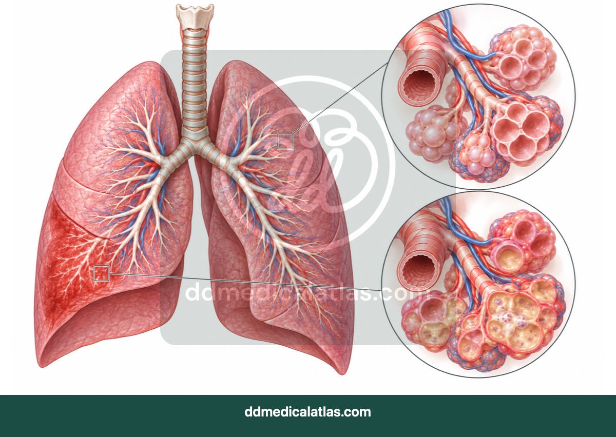
A medical illustration comparing healthy air-filled lung tissue with inflammatory fluid and consolidation associated with pneumonia.
Respiratory
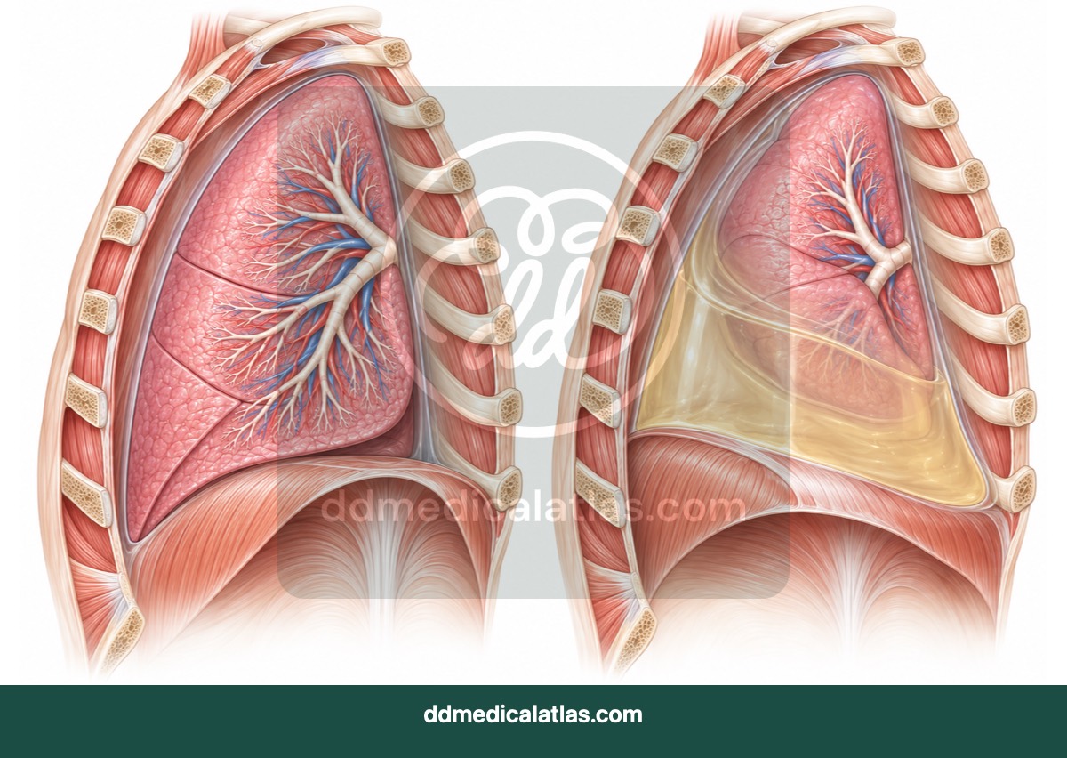
A medical illustration showing fluid accumulating in the pleural space and compressing the adjacent lung in pleural effusion.
Reproductive and breast health
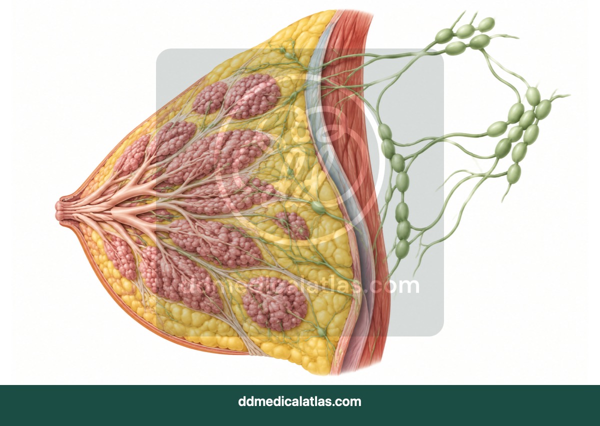
A medical illustration showing normal breast structure, glandular tissue, ducts and principal lymphatic drainage pathways.
Reproductive and breast health
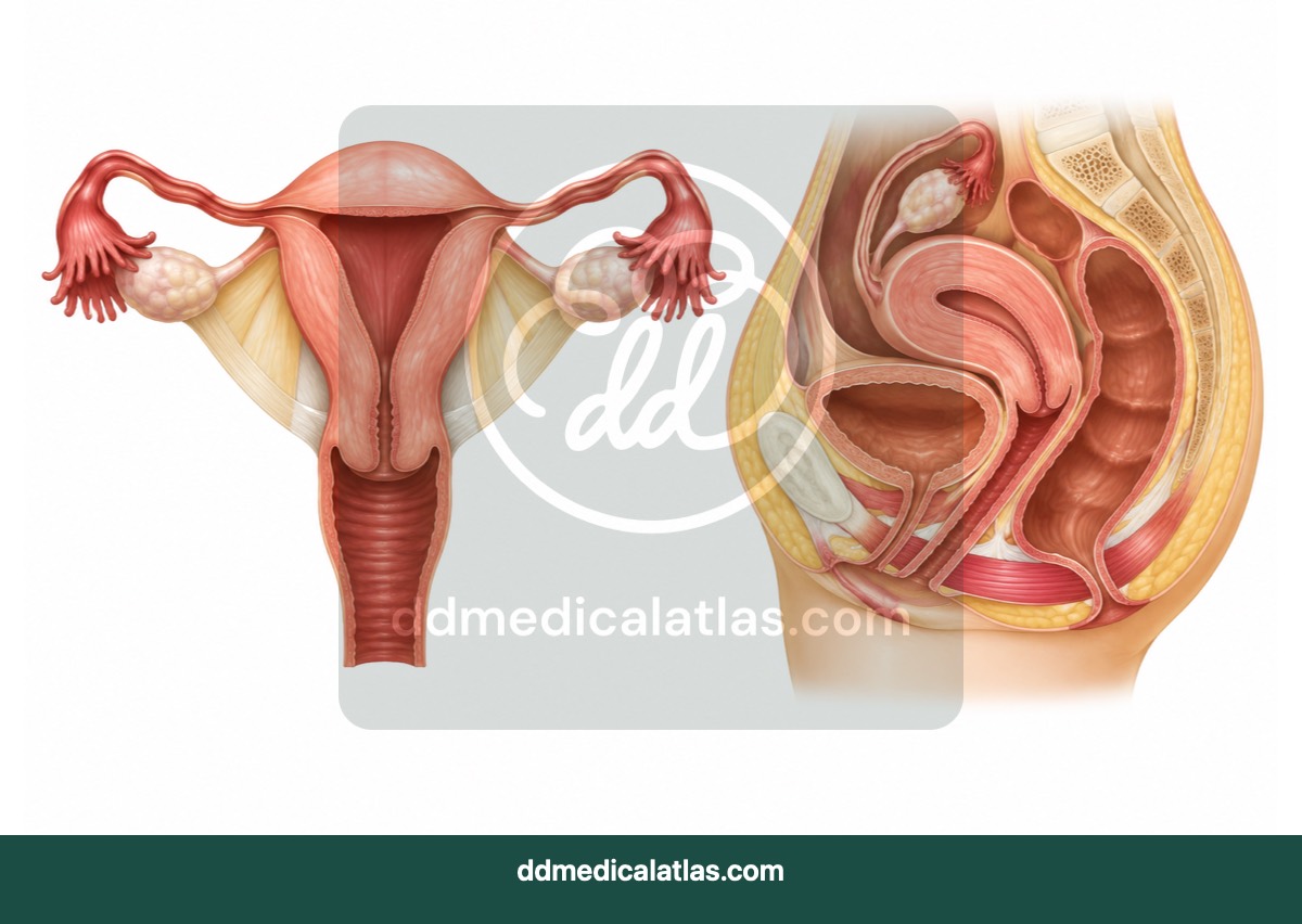
A medical illustration of the female reproductive system showing the uterus, ovaries, uterine tubes, cervix and vagina.